One of the most frustrating experiences for patients after a concussion is being told:
“Your MRI is normal.”
We want to see something on a test that validates how we feel, but following concussion, that validation doesn’t always come from traditional imaging. It comes from examining how the brain is actually functioning—through eye movements, balance, memory and cognition, proprioception, vestibular function, and autonomic regulation.
Meanwhile, patients don’t feel normal at all. They may still have headaches, brain fog, dizziness, overwhelming fatigue, difficulty reading, visual tracking problems, exercise intolerance, poor balance, memory changes, or feel spacey and disoriented. Driving or walking through a grocery store may suddenly feel overwhelming. They know their brain isn’t functioning the way it did before the injury, but their MRI says everything looks fine.
How can both things be true?
Because a traditional MRI is primarily looking at the structure of the brain. A concussion can significantly alter how the brain functions without creating the type of structural damage that a conventional MRI was designed to see.
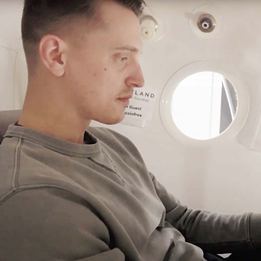
This distinction is also one of the reasons we use Hyperbaric Oxygen Therapy (HBOT) for patients recovering from concussion and traumatic brain injury. Advanced imaging research has demonstrated that brain injury can alter cerebral blood flow, metabolism, and neurological function even when conventional structural imaging doesn’t fully explain a patient’s symptoms. HBOT dramatically increases oxygen availability to brain tissue while research suggests it can also influence cerebral blood flow, cellular metabolism, angiogenesis, inflammation, and neuroplasticity.
In other words, we aren’t treating what the brain looks like on an MRI. We’re improving the biological environment the brain needs for healthy function and healing.
Structure and Function Are Not the Same Thing
A traditional MRI is an incredibly valuable tool. It can identify structural problems such as bleeding, tumors, significant tissue damage, swelling, and other anatomical abnormalities. After a head injury, ruling out these problems can be extremely important.
But concussion is often different.
A concussion can disrupt brain cells and their connections, alter mitochondrial energy production, change cerebral blood flow, affect autonomic regulation, increase inflammatory signaling, and change how different areas of the brain communicate with one another. Many of these changes occur at a physiological level rather than creating a large structural lesion that can easily be seen on conventional imaging.
The brain can look normal structurally while functioning very differently physiologically.
And research using more advanced brain-imaging techniques is helping us see this.
We Can Actually See Changes in Blood Flow After Concussion
The brain is incredibly dependent upon blood flow because blood delivers the oxygen and glucose our neurons need to produce energy. Although the brain makes up only about 2% of our body weight, it consumes roughly 20% of our body’s oxygen at rest.
Following concussion, however, the normal regulation of blood flow within the brain can become disrupted.
One important study published in JAMA Neurology used arterial spin labeling MRI, an advanced MRI technique that measures cerebral blood flow, to follow collegiate athletes after concussion. Researchers found evidence of reduced cerebral blood flow following concussion and subsequent recovery over time. Importantly, athletes who took longer to recover continued to demonstrate decreased blood flow within part of the insular cortex one month after their concussion.
Other studies using advanced perfusion MRI have also identified abnormal patterns of cerebral blood flow after traumatic brain injury. Interestingly, the pattern isn’t always simply “low blood flow everywhere.” Depending upon the injury and when imaging is performed, researchers have identified areas of both hypoperfusion and hyperperfusion.
That’s an important distinction.
The problem after brain injury may not simply be that the brain needs more blood. The problem may be that the brain has lost some of its ability to appropriately regulate where and when that blood needs to go.
And that matters tremendously because blood flow delivers oxygen. Oxygen allows mitochondria to produce ATP, and ATP provides the energy neurons require to communicate, maintain their electrical gradients, repair damaged structures, maintain synapses, and create the new connections involved in neuroplasticity.
Blood flow → Oxygen → Energy → Brain Function and Healing
When any part of that chain is disrupted, the brain may have a harder time functioning efficiently and repairing itself.
This Helps Explain Why You Can Feel So Bad With a “Normal” MRI
Your brain isn’t simply a collection of anatomical structures. It is an incredibly complex, interconnected network that is constantly processing and integrating information.
Reading, for example, requires coordinated eye movements, visual processing, attention, memory, language, vestibular stability, and cognitive processing. Driving requires even more. Your brain must simultaneously process speed, movement, peripheral vision, eye and head movements, balance, spatial orientation, attention, decision-making, and autonomic regulation.
Walking through a grocery store requires your brain to integrate:
- information from your eyes
- movement detected by your vestibular system
- sensory information from your muscles and joints
- head and neck position
- peripheral visual movement
- balance and spatial orientation
- attention and cognitive processing
Before your concussion, your brain may have performed all of this almost effortlessly. After concussion, communication between these systems may become less efficient while blood flow, oxygen utilization, and cellular energy production may also be altered.
That’s why patients frequently tell us:
“I can do it, but it takes so much more effort than it used to.”
Or:
“Everything looks normal, but I don’t feel like myself.”
They’re often describing dysfunction that a conventional structural MRI was never designed to measure.
This is where evaluating function becomes so important. Eye movements, vestibular function, balance, proprioception, memory and cognition, and autonomic regulation can provide information about how different neurological systems are actually performing and communicating with one another. Chiropractic/Functional Neurologists, vestibular-based Physical and Occupational Therapists, and Optometrists who specialize in neurological and visual rehabilitation can help identify these functional abnormalities and determine appropriate neuroplasticity-based treatment strategies.
Where Hyperbaric Oxygen Therapy Comes In
This is where Hyperbaric Oxygen Therapy becomes especially important in the conversation.
HBOT combines increased atmospheric pressure with high concentrations of oxygen, dramatically increasing the amount of oxygen dissolved directly into the plasma. This allows substantially more oxygen to circulate throughout the body and diffuse into tissues.
For the injured brain, that can be particularly meaningful because healing requires energy, energy production requires oxygen, and brain injury can disrupt the circulation and metabolism responsible for delivering and utilizing that oxygen.
Research in traumatic brain injury has directly measured some of these effects. In a prospective randomized clinical trial of patients with severe TBI, researchers directly measured oxygen levels within brain tissue as well as cerebral blood flow and oxygen metabolism. HBOT produced a dramatic increase in brain-tissue oxygen and significantly increased cerebral blood flow and cerebral oxygen metabolism following treatment.
The metabolic findings are particularly important. The goal isn’t simply to flood the brain with oxygen while someone is inside a hyperbaric chamber. The goal is to improve the biological environment in a way that allows injured brain tissue to use oxygen and produce the energy it needs for healthy function and repair.
Research in people with chronic post-concussion syndrome has also demonstrated changes in brain perfusion following a course of HBOT. In a randomized prospective crossover trial involving patients who continued to have symptoms one to five years after mild traumatic brain injury, researchers evaluated cognitive function along with SPECT brain-perfusion imaging before and after 40 HBOT sessions.
Following HBOT, patients demonstrated improvements in cognitive function and quality of life along with increased brain activity and perfusion on SPECT imaging. During the control period without HBOT, the same type of improvement was not observed.
This is particularly important because these weren’t people treated immediately after their concussion. They were still experiencing persistent problems years later, yet changes in brain function and perfusion were still measurable following treatment.
And the evidence that HBOT can improve cerebral circulation isn’t limited to concussion or traumatic brain injury.
In a 2021 study examining vascular dysfunction and aging, researchers used live imaging to follow individual cerebral blood vessels before and after HBOT. Following treatment, they found significant improvements in cerebral blood flow and changes in the blood vessels themselves. In the human portion of the study, older adults with significant memory loss also demonstrated increased cerebral blood flow along with improvements in cognitive performance following HBOT.
Other controlled human research has demonstrated similar changes. In healthy older adults, MRI perfusion imaging demonstrated significant regional increases in cerebral blood flow following a course of HBOT, particularly within frontal, parietal, and supplementary motor regions. These changes occurred alongside improvements in cognitive performance.
A randomized controlled trial in patients with post-COVID condition also demonstrated significant increases in cerebral blood flow on MRI perfusion imaging following HBOT compared with controls, including areas involved in attention, memory, sensory processing, and cognitive function.
These are obviously different patient populations, and we’re not suggesting that aging, Long COVID, and concussion are the same condition. What they help demonstrate is something more fundamental: repeated Hyperbaric Oxygen Therapy can create measurable changes in cerebral perfusion and the vascular environment of the brain.
HBOT Is About More Than Increasing Oxygen Today
Increasing oxygen availability is the immediate effect of Hyperbaric Oxygen Therapy, but it isn’t the entire reason we use it.
Repeated HBOT exposures can influence several biological processes involved in longer-term brain recovery, including:
- dramatically increasing tissue oxygen availability
- supporting mitochondrial energy production and cellular metabolism
- influencing cerebral circulation and oxygen utilization
- reducing inflammatory signaling
- stimulating angiogenesis—the growth and repair of blood vessels
- mobilizing stem and progenitor cells
- supporting neuroplasticity and regenerative signaling
Autonomic regulation may be another important part of this picture. The autonomic nervous system helps regulate heart rate, blood pressure, circulation, digestion, and many of the automatic processes required to maintain a healthy internal environment. Autonomic dysfunction is increasingly recognized following concussion and traumatic brain injury. Interestingly, human research by Lund et al has demonstrated that hyperbaric oxygen can increase parasympathetic activity, suggesting that HBOT may help influence the balance between sympathetic “fight-or-flight” activity and parasympathetic “rest-and-recovery” function. While this particular study was performed in professional divers rather than concussion patients, the finding is especially relevant because healthier autonomic regulation may support circulation, recovery, sleep, digestion, and overall brain-body function.
Angiogenesis is particularly important to this conversation.
If concussion or traumatic brain injury damages or disrupts the microvascular system responsible for delivering blood and oxygen to brain tissue, increasing oxygen during treatment helps provide more of what the brain needs immediately. But repairing and growing the vascular network may help improve the brain’s ability to deliver oxygen and nutrients long after the treatment itself is over.
Research into what’s known as the hyperoxic-hypoxic paradox helps us understand how this may occur. Repeated elevations in oxygen followed by a return toward normal oxygen levels can activate cellular signaling pathways involved in adaptation and repair—including pathways associated with angiogenesis, stem-cell mobilization, mitochondrial function, and neuroplasticity.
In other words, HBOT isn’t simply about giving the brain more oxygen for an hour.
We’re providing dramatically more oxygen today while stimulating biological processes that may improve the brain’s ability to deliver and utilize oxygen tomorrow.
This is one of the things that makes HBOT different. We’re trying to create an environment that supports the body’s own repair processes so that circulation, metabolism, cellular energy production, and neurological function can continue to improve.
A Normal MRI Is Good News—But It Isn’t the Whole Story
If your MRI is normal after concussion, that’s generally a good thing. It means the test did not identify the significant structural abnormalities it was designed to detect.
But it doesn’t necessarily mean your brain is functioning normally.
Concussion can affect blood flow, cellular metabolism, mitochondrial function, neurological signaling, autonomic regulation, inflammation, and communication between different areas of the brain without producing an obvious abnormality on a traditional MRI.
Advanced imaging research is increasingly allowing us to see some of these physiological changes, while functional neurological examination can help us identify how these changes are affecting the person sitting in front of us.
Hyperbaric Oxygen Therapy gives us a way to address several parts of that biological environment simultaneously by dramatically increasing oxygen availability while supporting circulation, energy metabolism, angiogenesis, inflammatory regulation, and neuroplasticity.
The brain doesn’t have to look damaged on an MRI to be struggling.
Understanding the mechanisms behind confusion that illustrate the difference between what the brain looks like and how the brain is actually functioning is the first step toward understanding why symptoms persist—and why we use hyperbaric oxygen therapy to help it heal.
Research
Meier TB, et al. Recovery of Cerebral Blood Flow Following Sports-Related Concussion. JAMA Neurology. 2015.
Wang Y, et al. Cerebral Blood Flow in Acute Concussion: Preliminary ASL Findings from the NCAA-DoD CARE Consortium. Brain Imaging and Behavior. 2019.
Rockswold SB, et al. A Prospective, Randomized Clinical Trial to Compare the Effect of Hyperbaric to Normobaric Hyperoxia on Cerebral Metabolism, Intracranial Pressure, and Oxygen Toxicity in Severe Traumatic Brain Injury. Journal of Neurosurgery. 2010.
Boussi-Gross R, et al. Hyperbaric Oxygen Therapy Can Improve Post Concussion Syndrome Years After Mild Traumatic Brain Injury—Randomized Prospective Trial. PLOS ONE. 2013.
Tal S, et al. Hyperbaric Oxygen Therapy Can Induce Angiogenesis and Regeneration of Nerve Fibers in Traumatic Brain Injury Patients. Frontiers in Human Neuroscience. 2017.
Shapira R, et al. Hyperbaric Oxygen Therapy Alleviates Vascular Dysfunction and Amyloid Burden in an Alzheimer’s Disease Mouse Model and in Elderly Patients. Aging. 2021.
Hadanny A, et al. Cognitive Enhancement of Healthy Older Adults Using Hyperbaric Oxygen: A Randomized Controlled Trial. Aging. 2020.
Zilberman-Itskovich S, et al. Hyperbaric Oxygen Therapy Improves Neurocognitive Functions and Symptoms of Post-COVID Condition: Randomized Controlled Trial. Scientific Reports. 2022.
Gottfried I, et al. Hyperbaric Oxygen Therapy as a Neuromodulatory Technique: A Review of the Recent Evidence.Frontiers in Neurology. 2024.
Lund V, Kentala E, Scheinin H, et al. Hyperbaric Oxygen Increases Parasympathetic Activity in Professional Divers. Acta Physiologica Scandinavica. 2000. PMID: 10971221.